Deep tissue super-resolution imaging with adaptive optical two-photon multifocal structured illumination microscopy
doi: 10.1186/s43074-023-00115-2
Deep tissue super-resolution imaging with adaptive optical two-photon multifocal structured illumination microscopy
-
Abstract:
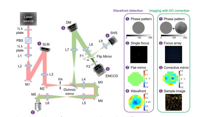
Visualization of axons and dendritic spines is crucial in neuroscience research. However, traditional microscopy is limited by diffraction-limited resolution and shallow imaging depth, making it difficult to study neuronal dynamics. Two-photon multifocal structured illumination microscopy (2P-MSIM) provides super-resolution imaging along with a reasonably good penetration, but it is vulnerable to optical aberrations in deep tissues. Herein we present a novel non-inertial scanning 2P-MSIM system incorporated with adaptive optics (AO) which allows for super-resolution imaging with effective aberration correction. Our strategy is designed to correct both laser and fluorescence paths simultaneously using a spatial light modulator and a deformable mirror respectively, providing better results than the individual path corrections. The successful implementation of adaptive optical two-photon multifocal structured illumination microscopy (AO 2P-MSIM) has allowed for the super-resolution imaging of neuronal structures in a mouse brain slice at great depths and dynamic morphological characteristics of zebrafish motoneurons in vivo.
-
Key words:
- Super-resolution /
- Adaptive optics /
- In vivo imaging /
- Neurons
-
[1] Al-Hasani R, Gowrishankar R, Schmitz GP, Pedersen CE, Marcus DJ, Shirley SE, et al. Ventral tegmental area GABAergic inhibition of cholinergic interneurons in the ventral nucleus accumbens shell promotes reward reinforcement. Nat Neurosci. 2021;24(10):1414–28. [2] Yuste R, Denk W. Dendritic spines as basic functional units of neuronal integration. Nature. 1995;375(6355):682–4. [3] Nimchinsky EA, Sabatini BL, Svoboda K. Structure and function of dendritic spines. Annu Rev Physiol. 2002;64:313–53. [4] Ji N, Shroff H, Zhong H, Betzig E. Advances in the speed and resolution of light microscopy. Curr Opin Neurobiol. 2008;18(6):605–16. [5] Turcotte R, Liang Y, Tanimoto M, Zhang Q, Li Z, Koyama M, et al. Dynamic super-resolution structured illumination imaging in the living brain. Proc Natl Acad Sci U S A. 2019;116(19):9586–91. [6] Gribble KD, Walker LJ, Saint-Amant L, Kuwada JY, Granato M. The synaptic receptor Lrp4 promotes peripheral nerve regeneration. Nat Commun. 2018;9(1):2389. [7] Denk W, Strickler JH, Webb WW. Two-photon laser scanning fluorescence microscopy. Science. 1990;248(4951):73–6. [8] Helmchen F, Denk W. Deep tissue two-photon microscopy. Nat Methods. 2005;2(12):932–40. [9] Ingaramo M, York AG, Wawrzusin P, Milberg O, Hong A, Weigert R, et al. Two-photon excitation improves multifocal structured illumination microscopy in thick scattering tissue. Proc Natl Acad Sci U S A. 2014;111(14):5254–9. [10] Winter PW, York AG, Nogare DD, Ingaramo M, Christensen R, Chitnis A, et al. Two-photon instant structured illumination microscopy improves the depth penetration of super-resolution imaging in thick scattering samples. Optica. 2014;1(3):181–91. [11] Zheng W, Wu Y, Winter P, Fischer R, Nogare DD, Hong A, et al. Adaptive optics improves multiphoton super-resolution imaging. Nat Methods. 2017;14(9):869–72. [12] Muller CB, Enderlein J. Image scanning microscopy. Phys Rev Lett. 2010;104(19): 198101. [13] Sheppard CJ, Mehta SB, Heintzmann R. Superresolution by image scanning microscopy using pixel reassignment. Opt Lett. 2013;38(15):2889–92. [14] Ji N, Milkie DE, Betzig E. Adaptive optics via pupil segmentation for high-resolution imaging in biological tissues. Nat Methods. 2010;7(2):141–7. [15] Sahu P, Mazumder N. Advances in adaptive optics-based two-photon fluorescence microscopy for brain imaging. Lasers Med Sci. 2020;35(2):317–28. [16] Ji N, Freeman J, Smith SL. Technologies for imaging neural activity in large volumes. Nat Neurosci. 2016;19(9):1154–64. [17] Booth M. Adaptive optical microscopy: the ongoing quest for a perfect image. Light Sci Appl. 2014;3(4):e165–165. [18] Zhou Z, Huang J, Li X, Gao X, Chen Z, Jiao Z, et al. Adaptive optical microscopy via virtual-imaging-assisted wavefront sensing for high-resolution tissue imaging. PhotoniX. 2022;3(1):1–20. [19] Shu Y, Sun J, Lyu J, Fan Y, Zhou N, Ye R, et al. Adaptive optical quantitative phase imaging based on annular illumination Fourier ptychographic microscopy. PhotoniX. 2022;3(1):24. [20] Wu J, Lu Z, Jiang D, Guo Y, Qiao H, Zhang Y, et al. Iterative tomography with digital adaptive optics permits hour-long intravital observation of 3D subcellular dynamics at millisecond scale. Cell. 2021;184(12):3318–32e17. [21] Tao X, Norton A, Kissel M, Azucena O, Kubby J. Adaptive optical two-photon microscopy using autofluorescent guide stars. Opt Lett. 2013;38(23):5075–8. [22] Wang K, Milkie DE, Saxena A, Engerer P, Misgeld T, Bronner ME, et al. Rapid adaptive optical recovery of optimal resolution over large volumes. Nat Methods. 2014;11(6):625–8. [23] Wang K, Sun W, Richie CT, Harvey BK, Betzig E, Ji N. Direct wavefront sensing for high-resolution in vivo imaging in scattering tissue. Nat Commun. 2015;6:7276. [24] Kashiwagi Y, Higashi T, Obashi K, Sato Y, Komiyama NH, Grant SGN, et al. Computational geometry analysis of dendritic spines by structured illumination microscopy. Nat Commun. 2019;10(1):1285. [25] Ke M-T, Nakai Y, Fujimoto S, Takayama R, Yoshida S, Kitajima Tomoya S, et al. Super-resolution Mapping of neuronal circuitry with an Index-Optimized Clearing Agent. Cell Rep. 2016;14(11):2718–32. [26] Balcioglu A, Gillani R, Doron M, Burnell K, Ku T, Erisir A, et al. Mapping thalamic innervation to individual L2/3 pyramidal neurons and modeling their ‘readout’ of visual input. Nat Neurosci. 2023;26(3):470–80. [27] Ruthazer ES, Li J, Cline HT. Stabilization of axon branch dynamics by synaptic maturation. J Neurosci. 2006;26(13):3594–603. [28] Niell CM, Meyer MP, Smith SJ. In vivo imaging of synapse formation on a growing dendritic arbor. Nat Neurosci. 2004;7(3):254–60. [29] Huang B, Li J, Yao B, Yang Z, Lam EY, Zhang J, et al. Enhancing image resolution of confocal fluorescence microscopy with deep learning. PhotoniX. 2023;4(1):1–22. [30] Liao J, Zhang C, Xu X, Zhou L, Yu B, Lin D, et al. Deep-MSIM: fast image reconstruction with deep learning in multifocal structured illumination microscopy. Adv Sci. 2023;10(27):2300947. [31] Kim D, Keesling A, Omran A, Levine H, Bernien H, Greiner M, et al. Large-scale uniform optical focus array generation with a phase spatial light modulator. Opt Lett. 2019;44(12):3178–81.
 下载:
下载:
 点击查看大图
计量
- 文章访问数: 258
- HTML全文浏览量: 3
- PDF下载量: 22
- 被引次数: 0